Research
Biomedical Optics
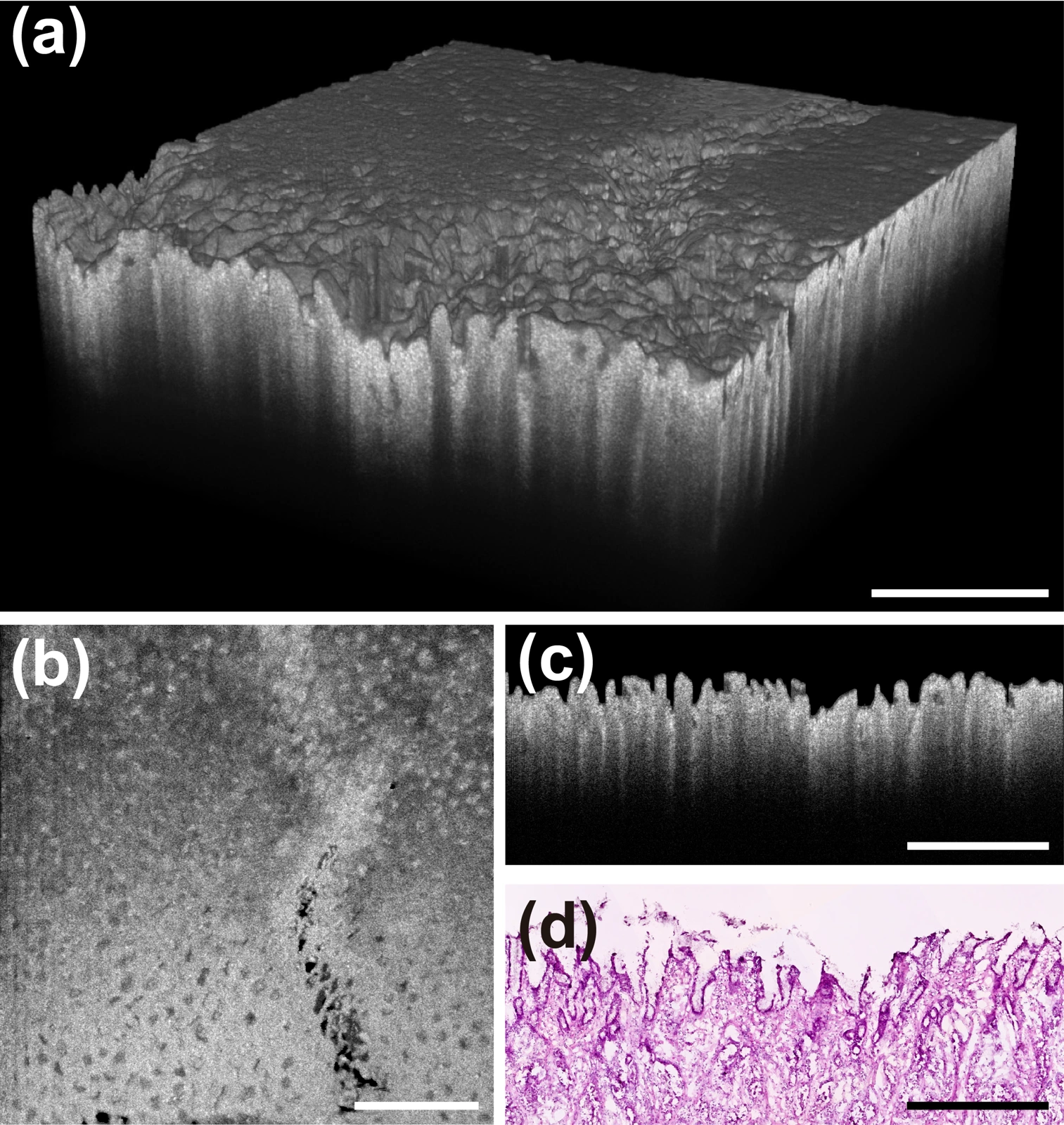
OCT & Catheter-based Imaging
Developing optical systems to reveal tissue structure and biochemical information.
We develop optical coherence tomography (OCT) systems and catheter-based imaging technologies to reveal tissue structure and complementary biochemical information. Our work spans benchtop instruments and miniature probes, combining optical design, scanning, and multimodal detection.
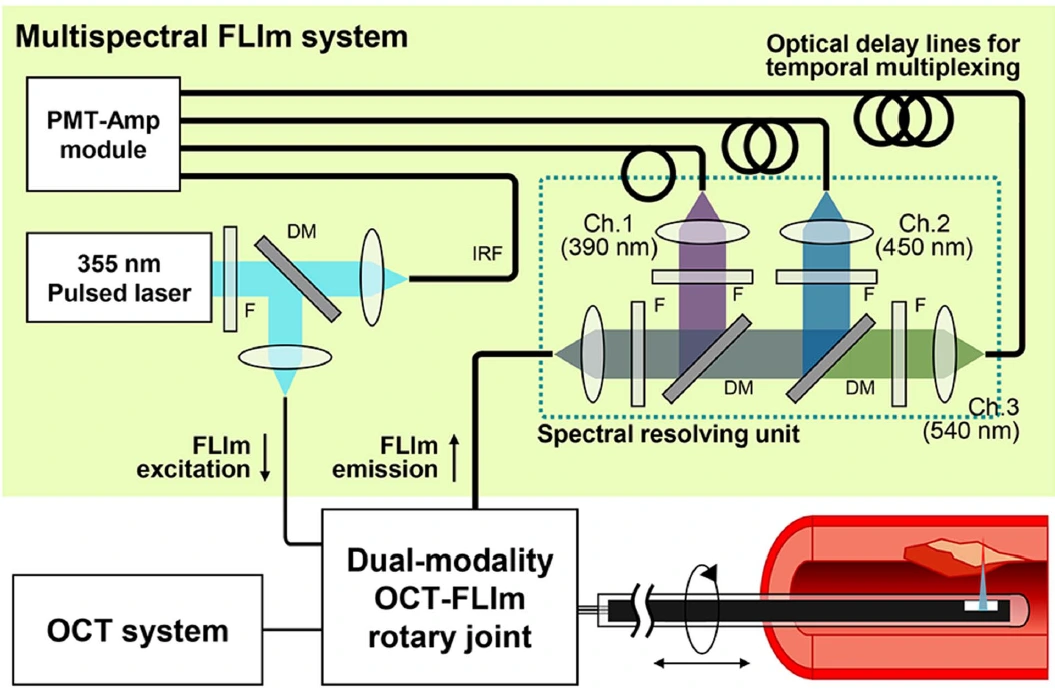
We design illumination, signal collection, and fiber-based interfaces to improve image quality and adapt imaging systems to different measurement settings. Integrating OCT with fluorescence lifetime imaging (OCT-FLIm) brings structural and biochemical information together, providing a platform for biomedical research and clinical imaging.
Cardiovascular Imaging & Theranostics
Understanding blood vessels beyond their shape.
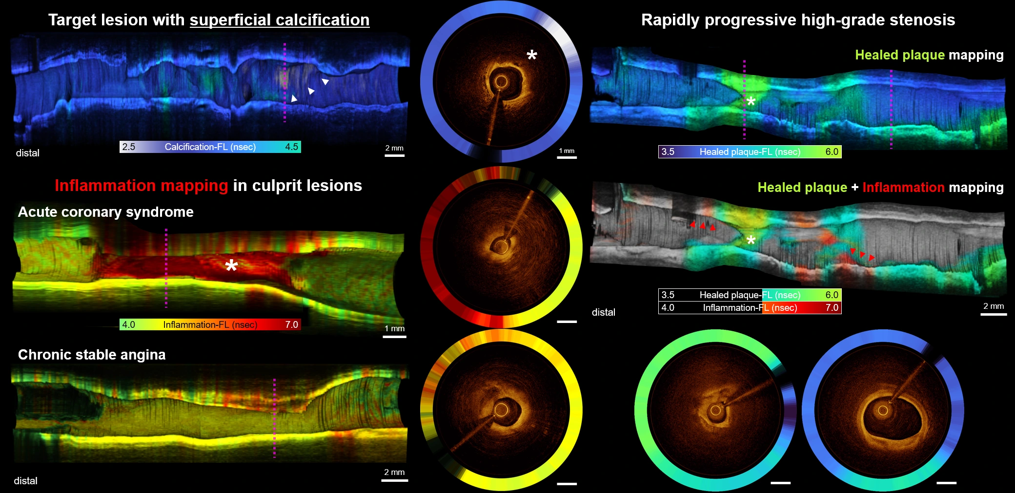
Understanding vascular disease requires information about both the structure of a blood vessel and the composition of its lesions. We develop intravascular OCT (IV-OCT) and combine OCT with fluorescence lifetime imaging (OCT-FLIm) or near-infrared fluorescence (NIRF) to explore these features together.
By linking detailed images with biochemical information, we aim to improve the assessment of atherosclerotic plaques. Alongside imaging, we investigate light-activated, targeted approaches to treatment, connecting the detection of disease with the study of therapeutic response.
Optical Biopsy & Label-free Imaging
Reading the information already present in tissue.
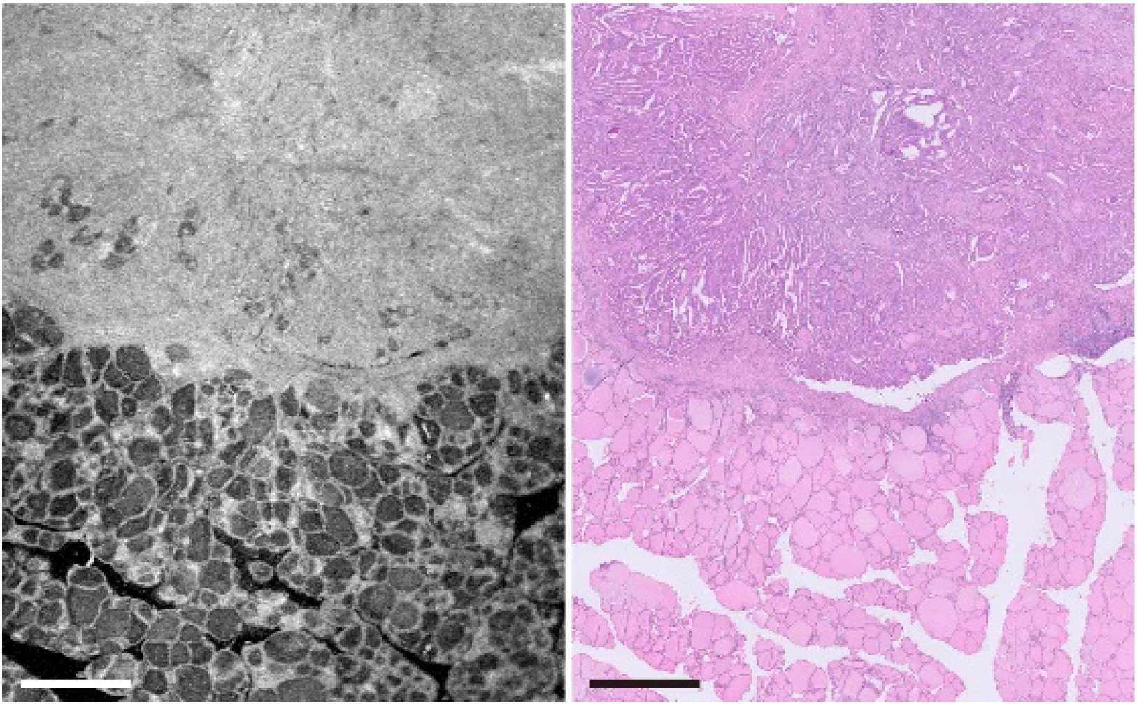
Tissue carries optical signatures that can reveal its structure and biochemical state. We use OCT, natural fluorescence, fluorescence lifetime imaging (FLIm), and Raman spectroscopy to explore these features without adding labels.
By combining optical images with quantitative analysis, we study how tissue changes with disease. Our research spans tissue specimens and biological models, building imaging approaches that can support tissue assessment and the understanding of biological processes.
Advanced 3D & Multimodal Microscopy
Seeing more of the structure and complexity of living systems.
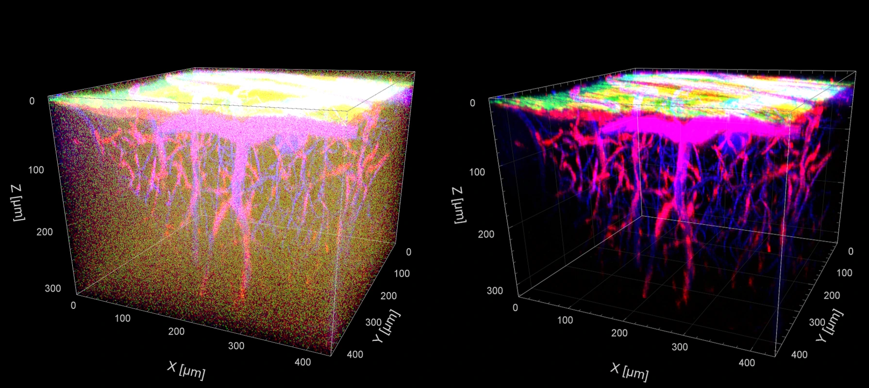
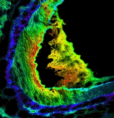
Cells and tissues are three-dimensional, and different imaging methods reveal different parts of their story. We develop multiphoton, light-sheet, and multimodal microscopes to bring these complementary views together.
By designing illumination and detection as an integrated system, we work to improve imaging speed, signal quality, and access to deeper structures. The result is a richer view of biological organization, from fine cellular features to larger tissue volumes.
AI for Optical Imaging
Turning complex optical data into clearer, more useful information.
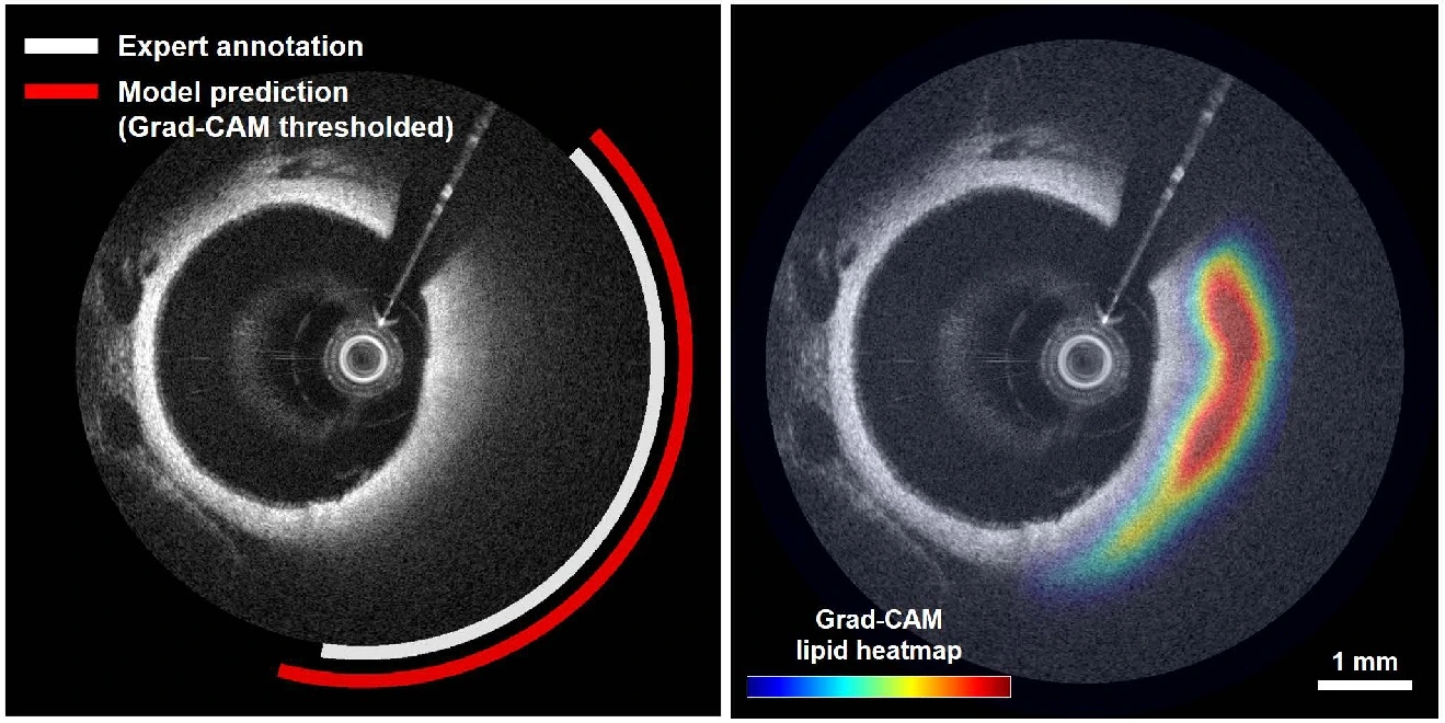
Weak signals, noise, and complex tissue structures can make optical images difficult to acquire and interpret. We develop AI methods that draw on the properties of optical signals and the spatial and temporal information within images.
Our research connects image restoration with quantitative analysis, from reducing noise while preserving fine structures to identifying tissue features and disease-related patterns. We aim to make optical imaging more informative for biological research and medical applications.